
MRI › Spine mri
Spine MRI in CypressTX for back and neck pain
A spine MRI maps discs, nerve roots and the spinal cord in detail no other study matches, without radiation. Cervical, thoracic and lumbar exams here are read by board certified radiologists.
30 to 45 minute scan
No radiation
Wide bore scanner
Results in 24 hours
What a spine MRI shows
Why MRI is the standard for spine complaints
X-ray reveals vertebrae and alignment but stops at bone. A spine MRI shows the soft structures causing most back and neck symptoms, including the discs themselves, the spinal cord running through the canal and every nerve root branching outward.
That is precisely why sciatica, numbness travelling down an arm or leg and weakness that will not resolve almost always lead to an MRI order rather than more films.
Conditions a spine MRI is ordered to investigate
What the scan itself is like
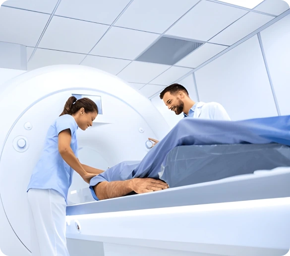
You lie flat on your back with a bolster placed beneath your knees, which flattens the lumbar curve and makes the position easier to hold. A flat coil sits under the region being imaged rather than around you, so nothing encloses the area being scanned.
Each sequence runs three to five minutes and the scanner knocks loudly while it works. Earplugs or headphones are provided and a squeeze bulb stays in your hand. Only the region ordered enters the scanner fully, so a lumbar study leaves your head near the opening.
Studies performed with contrast and without
Most spine MRI runs without any injection. Contrast becomes necessary when infection or tumor is suspected or when a patient has had previous spine surgery, because gadolinium is what separates scar tissue from a genuinely recurrent disc herniation on the images.
Spine MRI at a glance
Scan time
30 to 45 minutes
Time at the center
About 70 minutes
Radiation
None
Fasting
Not required
Referral
Required from your physician
Regions
Cervical thoracic and lumbar
Report turnaround
Typically 24 hours
Before your appointment
How to prepare for a Spine MRI
Very little is required in advance. Screening for implants and surgical hardware is the step that genuinely matters before a magnet this strong is switched on.
Report any spinal hardware
Rods, screws, cages and plates from previous surgery are usually MRI safe, though we need the details in advance. Spinal cord stimulators and pain pumps require specific settings and sometimes a representative present.
Eating and medicating normally
No fasting applies to spine imaging. Take pain medication before you arrive if that helps you lie flat comfortably, since staying still for forty minutes is the hardest part for many patients.
Choose clothing without metal
Soft pants and a plain shirt work best, avoiding zippers, studs and underwire. Belts and back braces come off before scanning. A gown and a locked cubby are available whenever you need them.
Bring prior films and reports
Earlier X-rays, CT scans or MRI discs let the radiologist judge whether a disc has worsened or held steady. That comparison often changes the recommendation your surgeon or pain specialist makes.
Other MRI studies
Related scans we perform
Ordered for a different region? Begin here instead.
Full Body MRI
Broad preventive survey across major organs
Cardiac MRI
Heart muscle, chamber volume and scar tissue
Musculoskeletal MRI
Ligament, tendon and cartilage injury
Brain MRI
Stroke lesions, tumors and MS plaques
Bone X-Ray
Fracture confirmation and healing checks
Spine X-Ray
Alignment, curvature and compression fractures
Common questions
Brain MRI questions patients ask
Book your spine MRI appointment
Send your referral over and we will complete safety screening, verify your coverage and hold a time. Our center sits at 18057 HWY 105 Suite 250 in Cypress, serving Conroe and Willis.