
MRI › Brain and head
Brain MRI scan serving Conroe and Cypress, TX
A brain MRI captures the finest detail obtainable of brain tissue, vessels and surrounding anatomy, with no radiation involved. Studies performed here are interpreted by a radiologist with neuroradiology training.
30 to 45 minute scan
No radiation
Wide bore scanner
Results in 24 hours
What a brain MRI shows
Clarity that other head imaging simply cannot deliver
Head CT is quick and outstanding for spotting fresh bleeding or a fractured skull. Brain MRI addresses an entirely different set of questions. Reading water content instead of density, it separates grey from white matter, exposes tiny lesions and captures changes CT overlooks completely.
That explains why a doctor investigating multiple sclerosis, a pituitary abnormality or the source of a headache that will not resolve reaches for MRI over CT.
Conditions a brain MRI is ordered to investigate
What the scan itself is like
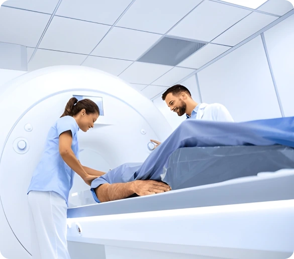
You rest on your back while a padded coil is arranged around your head and that coil captures the signal producing the image detail without ever touching your face. The table slides into the wide bore opening while your technologist remains in contact with you the whole time.
Expect loud bursts of noise as each sequence runs and you will receive earplugs or headphones beforehand. A call button stays in your hand throughout. Holding still is what matters most, since any movement blurs images and may force a sequence to be repeated.
With contrast or without
Certain brain exams include gadolinium delivered through an IV in your arm, which brightens tumors, inflammation and regions where the blood brain barrier has broken down. That call belongs to your ordering physician. When contrast is planned, kidney function is checked first and each step explained before we start.
Brain MRI at a glance
Scan time
30 to 45 minutes
Time at the center
About 75 minutes
Radiation
None
Fasting
Not required
Referral
Required from your physician
Read by
Neuroradiology trained
Report turnaround
Typically 24 hours
Before your appointment
How to prepare for a brain MRI
Almost nothing is asked of you beforehand. Safety screening is the exception that truly counts, since metal and a powerful magnetic field are incompatible.
Complete safety screening
Disclose any pacemaker, aneurysm clip, cochlear implant, stent, insulin pump or metal fragment lodged in the eye. Certain implants scan safely and others cannot, so this occurs before your slot is held.
Eat and take medication normally
Fasting is not required for brain MRI. Continue your prescriptions on their normal schedule unless your physician has specifically instructed you differently.
Leave metal and makeup out
Leave jewelry, hairpins and anything with zippers or underwire behind. Certain eye makeup and hair products carry metallic particles, so arrive with none of them applied.
Arrive fifteen minutes early
Bring your order, photo ID, insurance card and any earlier brain imaging on disc. A prior study gives your radiologist something to compare against, which usually strengthens the report considerably.
Other MRI studies
Related scans we perform
If your physician ordered a different body part, start here.
Spine MRI
Disc herniation, stenosis, nerve compression
Full Body MRI
Broad preventive survey across major organs
Cardiac MRI
Heart muscle, chamber volume and scar tissue
Musculoskeletal MRI
Ligament, tendon and cartilage injury
CT Without Contrast
Acute head bleeding and skull fracture
Vascular Ultrasound
Carotid narrowing and stroke risk assessment
Common questions
Brain MRI questions patients ask
Book your brain MRI appointment
Forward your referral and we will handle safety screening, confirm your coverage and secure a time. Find us at 7015 Barker Cypress Rd, Cypress,TX 77433, United States.