X-Ray › Chest X-ray
Chest X-ray with no appointment in Montgomery County
A chest X-ray remains the quickest way to assess the lungs, heart size and chest wall, finishing in minutes with minimal radiation. Board certified radiologists interpret every film.
About 10 minute visit
Walk-in accepted
Two standard views
Often read same day
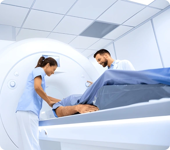
What a Chest X-ray shows
Lungs heart and airways captured in seconds
More chest X-rays are performed than any other imaging study and for good reason. In seconds it displays both lungs, the heart outline, the major airways, the diaphragm, the ribs and the soft tissues surrounding them.
It rarely gives a complete answer on its own, but it reliably tells the physician whether something is wrong and whether a CT scan is the necessary next step.
Conditions a chest X-ray commonly evaluates
What the scan itself is like
You stand facing a flat detector panel with your chest pressed lightly against it and your hands placed on your hips. The technologist asks you to take a deep breath in and hold it while the exposure is made.
A second image follows from the side. Each exposure takes a fraction of a second and the entire appointment is usually over within ten minutes of entering the room.
Why the breath hold matters
Fully inflated lungs spread out and become transparent to the beam, which is what makes abnormalities visible. A film taken mid breath compresses everything together and can create shadows resembling pneumonia where none exists.
Chest X-ray at a glance
Scan time
Under 10 minutes
Time at the center
Roughly 20 minutes
Radiation
Very low dose
Fasting
Not required
Referral
Required no appointment
Standard views
Front and side
Report turnaround
Typically 24 hours
Before your appointment
How to prepare for a chest X-ray
Nothing needs doing in advance beyond turning up with your order and clothing that will not interfere.
Remove metal from the chest area
Necklaces, underwire bras, shirt studs and medication patches all appear on the film. You will change into a gown if what you are wearing includes any of them.
Practise a full breath hold
A deep breath held steadily produces a far better image. Two or three practice breaths before the exposure make the difference between a clear film and one needing repeating.
Tell us about pregnancy
Chest films use very little radiation but the conversation is still worth having. Mention it at check in so shielding or an alternative approach can be arranged.
Bring earlier chest films
Comparison is often the whole story. A shadow present and unchanged for five years is reassuring, while the same shadow appearing for the first time warrants investigation.
Other MRI studies
Other scans available at our center
Need something more detailed? Start here.
Digital X-Ray
Walk in radiography for other body regions
Abdominal X-Ray
Bowel gas patterns and free air
Bone X-Ray
Rib fractures and skeletal assessment
Spine X-Ray
Coronary calcium scoring and plaque burden
High-Resolution CT Scan
Ultra thin slices for fine lung detail
Low Dose CT Lung Screening
Smokers annual screening
Common questions
Chest X-ray questions patients ask
Walk in for a chest X-ray
Bring your order any business day up to an hour before closing or call first and we will verify your benefits. We are at 18057 HWY 105 Suite 250 in Montgomery.